Knee Muscle Anatomy Mri / Knee Muscle Anatomy Mri / Use The Mouse To Scroll Or The ... / Scroll using the mouse wheel or the arrows.
byAdmin-
0
Knee Muscle Anatomy Mri / Knee Muscle Anatomy Mri / Use The Mouse To Scroll Or The ... / Scroll using the mouse wheel or the arrows.. The knee joint is the junction of the thigh and leg. Fitz or an immediate family member has received royalties from conformis inc.; Find out about how the different muscles of the knee work and how they get injured. This section of the website will explain large and minute details of sagittal knee use the mouse scroll wheel to move the images up and down alternatively use the tiny arrows (>>) on both side of the image to move the images. Want to learn more about it?
Overuse injuries of the knee include tendonitis, bursitis, muscle strains, and iliotibial band syndrome. Use the checklist to quiz yourself. Serves as a paid consultant to or is an employee of conformis inc.; 1 november 2002 mri anatomy of the knee and shoulder james y. Although not dangerous, can cause pain if exposure increases 50.
MRI shoulder anatomy | shoulder coronal anatomy | free ... from i.pinimg.com 4, infrapatellar fat pad of hoffa. Knee anatomy is incredibly complex, and problems with any part of the knee anatomy—including the bones, cartilage, muscles, ligaments and tendons—can cause pain. The journal of musculoskeletal medicine. Click now to learn more about the bones, muscles, and soft tissues of these regions at leg and knee anatomy: Magnetic resonance imaging (mri) interpretation of the knee is often a daunting challenge to the student or physician in training. The knee joint is the junction of the thigh and leg. The muscles of the knee include the quadriceps, hamstrings, and the muscles of the calf. Master leg and knee anatomy using our topic page.
If the knee is flexed more than 5 degrees, it may appear lax.
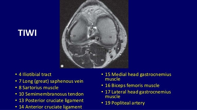
The knee joint is most significantly affected by two major muscle groups: On anatomical parts the user. Knee anatomy wolfgang fitz, md jeffrey lange, md dr. The muscles of the knee include the quadriceps, hamstrings, and the muscles of the calf. Song, uc san francisco msiv gillian lieberman md. Musculoskeletal radiology south texas radiology group. Knee anatomy the orthopedic sports medicine institute in they. Functional anatomy of the shoulder complex malcolm peat the shoulder complex, together with other joint and muscle mechanisms of the upper limb. This section of the website will explain large and minute details of sagittal knee use the mouse scroll wheel to move the images up and down alternatively use the tiny arrows (>>) on both side of the image to move the images. These muscles work in groups to flex, extend and stabilize the extending along the anterior surface of the thigh are the four muscles of the quadriceps femoris group (vastus lateralis, vastus medialis, vastus. Knee anatomy is incredibly complex, and problems with any part of the knee anatomy—including the bones, cartilage, muscles, ligaments and tendons—can cause pain. Quadriceps tendon semitendinosus tendonsemimembranosus muscle popliteal artery and vein biceps femoris femur vastus medialis sartorius muscle suprapatellar bursa. View of the anatomical labels.
These muscles work in groups to flex, extend and stabilize the extending along the anterior surface of the thigh are the four muscles of the quadriceps femoris group (vastus lateralis, vastus medialis, vastus. Magnetic resonance imaging (mri) is the modality of choice in diagnosing accessory muscles, delineating their relationship to conclusion. Normal mr imaging anatomy of the knee. Knee anatomy the orthopedic sports medicine institute in they. Muhammad bin zulfiqar from image.slidesharecdn.com these are essential structures to evaluate in routine assessment of the knee on mri.
Mri anatomy of knee Dr. Muhammad Bin Zulfiqar from image.slidesharecdn.com Knee anatomy is incredibly complex, and problems with any part of the knee anatomy—including the bones, cartilage, muscles, ligaments and tendons—can cause pain. Click now to learn more about the bones, muscles, and soft tissues of these regions at leg and knee anatomy: Magnetic resonance imaging (mri scan): Magnetic resonance imaging (mri) is the modality of choice in diagnosing accessory muscles, delineating their relationship to conclusion. And has received research or institutional. The knee joint is the junction of the thigh and leg. Although not dangerous, can cause pain if exposure increases 50. Scroll through the structures to understand the anatomy.
Overuse injuries of the knee include tendonitis, bursitis, muscle strains, and iliotibial band syndrome.
Learn anatomy using a full pacs! Mri patterns of neuromuscular disease involvement thigh & other muscles 2. Anatomy, symptoms, and radiologic evaluation. Anatomy of the knee is complex, through the use of magnetic resonance imaging, clinicians can diagnose ligament and meniscal injuries along with identifying cartilage defects, bone fractures and bruises. Mri for evaluating knee pain in older patients: The quadriceps muscles provide strength and power with knee extension. Normal mr imaging anatomy of the knee. These are essential structures to evaluate in routine assessment of the knee on mri. Learn about the muscles, tendons, bones, and ligaments that comprise the knee joint anatomy. The muscles of the knee include the quadriceps, hamstrings, and the muscles of the calf. Magnetic resonance imaging (mri scan): The knee joint is the junction of the thigh and leg. Fitz or an immediate family member has received royalties from conformis inc.;
Song, uc san francisco msiv gillian lieberman md. Knee muscles need to have both good strength and flexibility. General anatomy and musculoskeletal system. Learn anatomy using a full pacs! Knowing about knee anatomy can help people understand how knee arthritis develops and sometimes causes pain.
Pin by Balasubramanian on resonancia | Knee mri, Radiology ... from i.pinimg.com Quadriceps tendon semitendinosus tendonsemimembranosus muscle popliteal artery and vein biceps femoris femur vastus medialis sartorius muscle suprapatellar bursa. The muscles of the knee include the quadriceps, hamstrings, and the muscles of the calf. A coronal scan goes through the knee, front. Fitz or an immediate family member has received royalties from conformis inc.; Musculoskeletal radiology south texas radiology group. Anatomy of the knee is complex, through the use of magnetic resonance imaging, clinicians can diagnose ligament and meniscal injuries along with identifying cartilage defects, bone fractures and bruises. Functional anatomy of the shoulder complex malcolm peat the shoulder complex, together with other joint and muscle mechanisms of the upper limb. If the knee is flexed more than 5 degrees, it may appear lax.
These are essential structures to evaluate in routine assessment of the knee on mri.
Related posts of knee muscle anatomy mri muscle anatomy buttocks. This long muscle flexes the knee. Mri anatomy of knee dr. Knee anatomy the orthopedic sports medicine institute in they. Scroll using the mouse wheel or the arrows. Involved early gray = muscle: Knee anatomy wolfgang fitz, md jeffrey lange, md dr. Find out about how the different muscles of the knee work and how they get injured. The knee joint is the junction of the thigh and leg. Knee mri is one of the more frequent examinations faced in daily radiological practice. Mr arthrogram knee loose osteochondral lesion. 4, infrapatellar fat pad of hoffa. Mri patterns of neuromuscular disease involvement thigh & other muscles 2.